I’m really nothing more than a photographer. I record what I see.
Jean-Marie Charcot, 7 February 1888
Hi Francesco, tell us a bit about yourself
Hi Ben, thank you for inviting me to your blog! I was born and raised in Udine, a small town in the north-eastern part of Italy, but I currently live in Modena and work in Bologna, at the IRCCS Istituto delle Scienze Neurologiche (Institute of Neurological Sciences). I started off my medical career as a full-time clinician, working my way up through many locums around Italy. It was a hard training but it gave me a chance to strengthen my competences in neurology by applying them to many other medical fields, from rehabilitation to ER, and to experience relationships with patients in diverse settings. I have been interested in epidemiology since my graduation in medical school, and about twenty years ago I made a big change in my career, devoting myself to epidemiology and the methodology of medical research while narrowing the time of spent on clinical practice (though I still do one day a week, since I think that even in a research setting it is important to see patients and listen to them).
Want some local neuro-history from Bologna?


My other great interest is in photography, that I have pursued since I was a teenager. I started taking pictures and building my own darkroom in the 1970’s, when photography was entirely analogue. I am self-taught, although I read many books and have attended workshops on various technical and historical topics about photography. My artistic production is often influenced by the relationships between photography and science. I am particularly fascinated by taxonomy, the effort of finding a name for each manifestation of nature, often based on its shape, which photography allows the representation of so accurately.
My interest in Cochrane came in the 1990’s when I was lucky enough to meet and work with Alessandro Liberati. He was an extraordinary person. His energy and enthusiasm were contagious and ten years after he passed I still feel every day his strong legacy. Currently I am co-ordinating editor of the Cochrane Review Group on Multiple Sclerosis and Rare Diseases of the CNS.
You work in neurology and have a passion for photography, as well an interest in the history of both. Is there overlap between these areas of interest?
Definitely. In the second half of the 19th century photography, among other effects, opened up unprecedented documentary possibilities in the scientific world. In fact, photography was announced as a new invention on January 6, 1839, at the French Academy of Sciences. For the first time it was possible to capture a detailed, reproducible image. It made it possible to compare images of the same individual over time or between individuals with similar features. Also, because it was carried out by a machine and produced a direct trace on a physical medium it was considered to be much more ‘objective’ and reliable than, for example, hand-drawn sketches.
Can you tell us about some of the ways photography influenced science and medicine?
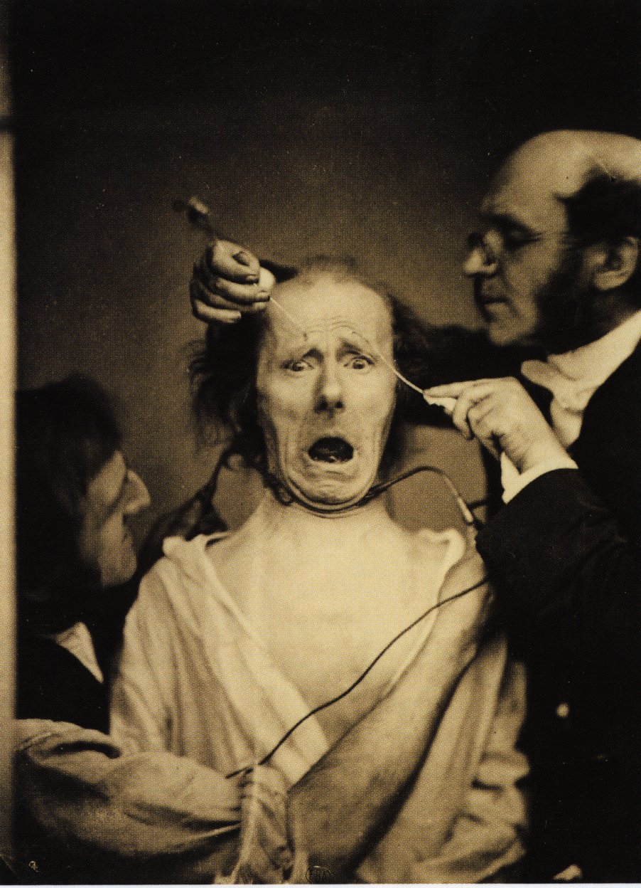
The first to systematically use photography in the medical field was a neurologist, Guillaume Duchenne de Boulogne (1806-1875). Duchenne published ‘Mécanisme de la physionomie humaine’, including a series of photographs (taken by Adrien Tournachon, better known as the brother of Nadar, 19th Century photography pioneer) that document Duchenne’s studies of the mimic or facial muscles which are responsible for making expressions. They feature a patient suffering from a peculiar insensitivity of the face, allowing Duchenne to apply metal electrodes to the patient’s skin and electrically stimulate specific muscles. Duchenne’s work aroused great interest among scientists of the time, including Charles Darwin. In 1872 Darwin published ‘The Expression of the Emotions in Man and Animals‘ in which the expressions of humans and primates are analysed and compared, in the context of his evolutionary theories. It included photographs by Duchenne and others.